Editorial Review
Author: PurePep Vital Research Editorial Team|Reviewed by: Scientific Compliance Reviewer
Last reviewed: February 2026
Innate vs Adaptive: Where Peptides Intervene
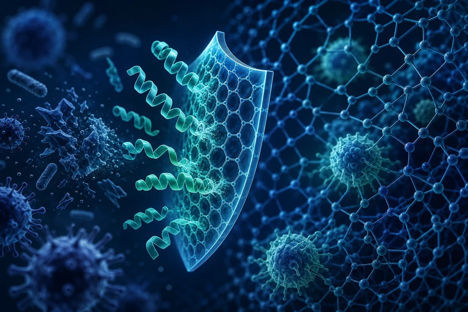
The immune system has two main branches. Innate immunity acts fast but broadly — it attacks any threat it finds. Adaptive immunity is slower but precise — it targets specific invaders and remembers them for next time.
Both branches rely on peptide signals to activate, regulate, and coordinate their responses. Cytokines, chemokines, antimicrobial peptides, and thymic hormones are all peptide-based molecules. They orchestrate immune function at every level.
Research on peptides for immune system modulation spans several categories:
- Immune-stimulating peptides that enhance defense capacity (thymosin alpha-1, LL-37)
- Immune-regulating peptides that prevent excessive inflammation (KPV)
- Tissue-repair peptides that support immune recovery after injury or infection (BPC-157, TB-500)
These distinctions matter. Immune modulation is bidirectional — the goal in research is rarely to simply "boost" immunity. Instead, researchers aim to restore the right immune function for a specific context.
The clinical significance of peptide immune modulation is clear. Thymosin alpha-1 holds approval as a therapeutic agent in over 35 countries for hepatitis B, hepatitis C, and as an immune-modulating adjunct. This makes it one of the few immune peptides with clinical validation beyond preclinical research. For foundational peptide biology, see our comprehensive peptide guide.
Thymosin Alpha-1: FDA Orphan Drug for Hepatitis B
Thymosin alpha-1 (Tα1) is a 28-amino-acid peptide that the thymus gland naturally produces. Dr. Allan Goldstein first isolated it at George Washington University School of Medicine in the 1970s.
Since then, Tα1 has become the most clinically validated immune-modulating peptide in existence. It holds FDA orphan drug designation and marketing approval in over 35 countries under the trade name Zadaxin.
Mechanisms of Immune Modulation
Thymosin alpha-1 enhances immune function through multiple mechanisms:
- It activates dendritic cells — immune cells that present threats to T cells — via Toll-like receptor 9 (TLR9) signaling. This bridges innate and adaptive immunity.
- It stimulates T-cell maturation in the thymus and beyond, restoring T-cell populations depleted by aging, infection, or immunosuppressive therapy.
- It boosts natural killer (NK) cell activity against virally infected and malignant cells.
- It shifts cytokine production toward Th1 (cell-mediated) immunity while keeping excessive inflammatory responses in check.
Clinical Evidence
Over 80 clinical trials have evaluated thymosin alpha-1 across diverse immune applications.
In chronic hepatitis B, a meta-analysis published in Journal of Viral Hepatitis showed that Tα1 monotherapy produced sustained virological response rates of 36–40%. These rates match interferon-alpha but come with far fewer side effects.
In hepatitis C, adding Tα1 to standard pegylated interferon/ribavirin therapy improved sustained response rates by about 15%.
Cancer Immunotherapy Adjunct
Researchers have evaluated Tα1 as an immunotherapy adjunct in multiple cancer types. A review in Expert Opinion on Biological Therapy found that Tα1 enhanced chemotherapy efficacy while reducing complications from immunosuppression.
In hepatocellular carcinoma, melanoma, and non-small cell lung cancer trials, Tα1 improved key immune markers (CD4/CD8 ratios, NK cell activity). It also reduced infection rates in immunocompromised study participants.
LL-37: Activity Against Resistant Bacteria
LL-37 is the only cathelicidin — a type of natural germ-fighting peptide — produced in the human body. It serves as a frontline component of innate immunity.
Neutrophils, macrophages, epithelial cells, and keratinocytes all produce LL-37. It provides immediate antimicrobial defense while also shaping broader immune responses.
Antimicrobial Spectrum
LL-37 kills pathogens directly by disrupting their cell membranes. Research published in Nature Reviews Microbiology documents activity against a wide range of threats:
- Gram-positive bacteria (MRSA, VRE)
- Gram-negative bacteria (E. coli, P. aeruginosa, K. pneumoniae)
- Mycobacteria (M. tuberculosis)
- Fungi (Candida species)
- Enveloped viruses (influenza, HIV)
This broad spectrum makes LL-37 especially valuable for studying antibiotic-resistant infections where standard drugs have failed.
Immune Cell Recruitment and Activation
Beyond direct killing, LL-37 acts as an immune alarm signal. It draws neutrophils, monocytes, and T cells to infection sites as a chemoattractant — a chemical that recruits immune reinforcements.
LL-37 also activates the FPRL1 receptor on immune cells. This triggers signaling cascades that enhance phagocytosis (pathogen engulfing), cytokine production, and antigen presentation. Research in Journal of Immunology shows that LL-37 bridges innate and adaptive immunity by promoting dendritic cell maturation and boosting antigen-specific T-cell responses.
Anti-Endotoxin Activity
LL-37 binds and neutralizes lipopolysaccharide (LPS) — a toxic molecule from gram-negative bacteria also called endotoxin. This matters greatly for sepsis research, where excessive endotoxin-driven inflammation (cytokine storm) is a leading cause of death.
Studies in Critical Care Medicine found that LL-37 reduced endotoxin-induced inflammatory mediator production by 60–80% in macrophage cultures. See our LL-37 research guide for comprehensive data.
Free Peptide Calculator
Calculate precise reconstitution volumes and dosages with our peptide calculator tool.
KPV: The Alpha-MSH Fragment Suppressing NF-kB
KPV (Lys-Pro-Val) is a tripeptide that addresses the regulatory side of immune function. It works to prevent excessive inflammation — the kind that damages host tissues and fuels autoimmune processes.
Immune-stimulating peptides like thymosin alpha-1 and LL-37 enhance defense capacity. KPV takes a different approach. It modulates inflammatory responses that, when out of control, drive autoimmune disease, chronic inflammation, and tissue damage.
NF-κB as an Immune Regulatory Target
NF-κB is a master transcription factor — a protein that switches genes on or off. It controls the expression of over 400 genes tied to immune and inflammatory responses.
In autoimmune conditions like rheumatoid arthritis, inflammatory bowel disease, psoriasis, and multiple sclerosis, NF-κB stays overactive. This drives persistent inflammation that damages host tissues.
KPV blocks NF-κB from entering the cell nucleus. This reduces production of key inflammatory mediators — TNF-α, IL-1β, IL-6, IL-8, and COX-2 — without shutting down the immune system broadly.
Autoimmune Disease Research
Research on α-MSH-derived peptides (KPV's parent molecule) in autoimmune models shows significant anti-inflammatory effects. Key findings include:
- In experimental autoimmune encephalomyelitis — a model for multiple sclerosis — melanocortin peptides reduced demyelination, lowered inflammatory cell infiltration into the CNS, and improved clinical scores.
- In collagen-induced arthritis models, α-MSH fragment treatment reduced joint inflammation and cartilage destruction.
These results suggest KPV may counter the inappropriate immune activation that underlies autoimmune pathology.
Selective Immune Modulation
KPV holds a key advantage over broad immunosuppressants like methotrexate, cyclosporine, and corticosteroids: selectivity. KPV suppresses NF-κB-driven inflammatory cascades without broadly shutting down immune function.
This means it may reduce autoimmune inflammation while preserving the ability to fight infections. Preclinical data supports this — KPV-treated animals maintained pathogen clearance despite showing reduced inflammatory markers. Read more in our KPV peptide guide.
Get Peptide Research Updates
New research, product launches, and exclusive offers. No spam.
BPC-157 and Gut-Associated Lymphoid Tissue
BPC-157 (Body Protection Compound-157) is best known for tissue repair. However, its effects on the immune-gut axis make it relevant to immune research too. This axis refers to the two-way communication between gut-associated lymphoid tissue (GALT) and the rest of the immune system.
GALT and Systemic Immunity
About 70–80% of the body's immune cells live in the gastrointestinal tract. This makes the gut the largest immune organ.
Gut-associated lymphoid tissue includes Peyer's patches, mesenteric lymph nodes, and a vast network of immune cells in the gut lining. BPC-157 directly influences this immune compartment through its effects on gut barrier integrity, mucosal healing, and enteric inflammation.
Immune-Relevant Mechanisms
Research published in Current Pharmaceutical Design shows that BPC-157 modulates several immune-relevant pathways:
- It stabilizes the nitric oxide (NO) system, which plays key roles in macrophage antimicrobial activity and vascular immune responses.
- It strengthens gut barrier integrity, reducing the leak of bacterial endotoxins that drive systemic inflammation.
- It modulates serotonin and dopamine systems, which influence immune cells through neuro-immune interactions.
- It reduces inflammation through prostaglandin and cytokine modulation.
Implications for Autoimmune Conditions
The immune-gut axis is gaining recognition as a driver of systemic autoimmunity. "Leaky gut" — increased intestinal permeability — allows bacterial antigens to cross into the bloodstream. This has been linked to the development of rheumatoid arthritis, type 1 diabetes, lupus, and multiple sclerosis.
BPC-157's ability to restore gut barrier function may carry systemic immune benefits beyond the GI tract. Explore the full BPC-157 research profile in our BPC-157 guide.
Important Disclaimer
All products and information on this page are intended strictly for laboratory and scientific research use only. Not for human consumption. These statements have not been evaluated by the FDA.
TB-500 in Wound-Mediated Immune Response
TB-500 is a synthetic fragment of thymosin beta-4 (Tβ4), a 43-amino-acid protein. Despite sharing the "thymosin" name with thymosin alpha-1, it works through entirely different mechanisms.
Tα1 is a thymic hormone that shapes T-cell development. Tβ4, by contrast, is a widespread intracellular protein involved in actin polymerization — the process cells use to move and change shape. It also supports cell migration and tissue repair. TB-500 has shown immune-relevant properties worth examining.
Anti-Inflammatory Mechanisms
TB-500 reduces inflammation through multiple pathways:
- Suppression of NF-κB activation
- Promotion of M2 (anti-inflammatory) macrophage polarization
- Reduction of pro-inflammatory cytokine production
In corneal alkali burn models, TB-500 treatment reduced inflammatory cell infiltration. It also promoted anti-inflammatory cytokine profiles that supported tissue regeneration. Research in Annals of the New York Academy of Sciences confirmed these effects across multiple tissue types.
Wound Healing and Immune Recovery
TB-500 promotes cell migration, new blood vessel formation (angiogenesis), and tissue remodeling. These actions support immune recovery after injury or infection by rebuilding the tissue structure that immune cells need to travel and patrol.
In sepsis models, thymosin beta-4 treatment improved survival rates and reduced organ damage. This suggests protective effects during overwhelming systemic immune challenges.
Immune Cell Migration
TB-500 regulates actin polymerization, which gives it influence over immune cell motility — the ability of immune cells to migrate toward infection or injury sites (chemotaxis).
Research shows that thymosin beta-4 promotes macrophage and lymphocyte migration. This may enhance immune surveillance and response to localized threats. The mechanism complements thymosin alpha-1: Tα1 activates immune cells while TB-500 helps deploy them where needed. See our TB-500 research guide for detailed data.
Comparing Immune Peptides by Pathway Target
The diversity of immune-modulating peptides enables targeted research for different immune contexts:
Immune Enhancement (Immunocompromised States): Thymosin alpha-1 is the gold standard for immune enhancement. Clinical evidence supports its use in chronic viral infections, cancer-related immunosuppression, and post-surgical immune recovery. LL-37 complements Tα1 by providing direct antimicrobial defense at barrier surfaces — skin, respiratory lining, and gut mucosa — where infections start.
Immune Regulation (Autoimmune States): KPV's selective NF-κB suppression targets the inappropriate immune activation behind autoimmune inflammation. It does this without broadly compromising immune function. BPC-157's gut barrier effects may reduce the antigenic stimulation from intestinal translocation that fuels systemic autoimmunity.
Immune Recovery (Post-Infection/Injury): TB-500 promotes tissue repair and anti-inflammatory macrophage polarization. This supports recovery of the tissue architecture needed for normal immune function. Combined with BPC-157 for wound healing and Tα1 for T-cell rebuilding, a comprehensive immune recovery approach emerges.
Combination Considerations: Multi-peptide immune protocols must distinguish between immune stimulation and immune regulation. Combining Tα1 (enhancement) with KPV (regulation) could theoretically boost pathogen defense while preventing inflammatory overshoot. However, such combinations require careful preclinical validation.
All immune modulation research should be conducted under appropriate institutional oversight. For broader context, see our peptide therapy overview.
| Peptide | Primary Pathway | Immune Arm | Key Model |
|---|---|---|---|
| Thymosin Alpha-1 | TLR / dendritic cell | Adaptive | Hepatitis B, cancer |
| LL-37 | Membrane disruption | Innate | Bacterial infection |
| KPV | NF-kB suppression | Regulatory | Colitis, inflammation |
| BPC-157 | VEGF / NO pathway | Gut-associated | IBD models |
| TB-500 | Actin regulation | Wound-immune | Tissue repair |
Research Limitations and Open Questions
Immune peptide research continues to expand, driven by advances in immunology, drug delivery, and precision medicine.
Checkpoint Immunotherapy Adjuncts: Researchers are evaluating thymosin alpha-1 alongside immune checkpoint inhibitors — drugs like anti-PD-1 and anti-CTLA-4 that unleash T cells against cancer. The rationale: Tα1 enhances T-cell populations, potentially improving the immune foundation these inhibitors rely on.
Early clinical data in hepatocellular carcinoma and non-small cell lung cancer shows improved response rates and fewer immune-related adverse events when Tα1 is added.
Antimicrobial Peptide Engineering: Synthetic biology now enables the design of antimicrobial peptides with better potency, selectivity, and stability. LL-37-derived analogs with improved protease resistance and lower host cell toxicity are advancing through preclinical development. These engineered peptides may help address antibiotic-resistant infections — a critical public health threat.
Immune Aging (Immunosenescence): Thymic function declines with age — a process called thymic involution. This leads to progressive immune dysfunction: reduced T-cell diversity, impaired vaccine responses, and increased infection susceptibility.
Thymosin alpha-1 research in elderly populations shows partial restoration of T-cell parameters. This suggests potential applications for age-related immune decline. Combined with telomere-focused interventions (epitalon), immune peptides may address multiple hallmarks of immune aging at once.
Vaccine Adjuvants: Both Tα1 and LL-37 demonstrate vaccine adjuvant properties. They enhance antibody responses and T-cell memory when co-administered with vaccines. This application is especially relevant for elderly populations, where immunosenescence reduces vaccine efficacy. Explore additional immune-relevant peptide research in our LL-37 guide and KPV overview.
Important Disclaimer — For Research Use Only
The information provided is for educational and research purposes only. All peptides discussed or linked on this site are intended strictly for laboratory and scientific research use only (RUO) and are not for human consumption, injection, ingestion, or any therapeutic application. These products have not been evaluated or approved by the FDA or any regulatory body and are not intended to diagnose, treat, cure, or prevent any disease or condition. Reliance on this content is at your own risk. Consult qualified professionals for any health-related decisions. PurePep Vital disclaims all liability for misuse. Products are offered by third-party retailers for research use only.
PurePep Vital is a chemical supplier. PurePep Vital is not a compounding pharmacy or chemical compounding facility as defined under 503A of the Federal Food, Drug, and Cosmetic Act. PurePep Vital is not an outsourcing facility as defined under 503B of the Federal Food, Drug, and Cosmetic Act.
Disclosure: This page contains affiliate links. We may earn from qualifying purchases. See our full disclosure.
Need deals after the research?
Use the deals hub to compare current offers, partner codes, and research news. We recommend retailers that provide quality signals when available.
Explore Our Guides
Related Articles
LL-37 Peptide: Antimicrobial Defense Research
LL-37 is the only cathelicidin-derived antimicrobial peptide in humans. Here is what research reveals about its role in innate immunity, wound healing, and infection defense.
KPV Peptide Benefits: Anti-Inflammatory Powerhouse Explained
KPV peptide is a potent anti-inflammatory derived from alpha-MSH. Here is what the research shows about its benefits for gut health, skin, and immune function.

TB-500 Peptide: Tissue Repair and Thymosin Beta-4
TB-500 is a synthetic fragment of thymosin beta-4, a naturally occurring 43-amino-acid protein involved in cell migration, differentiation, and tissue repair. Here is what the research reveals.
BPC-157 Peptide: The Complete Healing Research Guide
BPC-157 is a pentadecapeptide derived from human gastric juice with extraordinary tissue-healing properties. Here is what decades of research reveal about its mechanisms, benefits, and applications.
Frequently Asked Questions
Thymosin alpha-1 (Tα1) has the most extensive clinical validation, with over 80 clinical trials, FDA orphan drug designation, and marketing approval in 35+ countries. It enhances dendritic cell activation, T-cell maturation, NK cell cytotoxicity, and Th1 immunity. Clinical evidence supports its use in chronic hepatitis B (36-40% sustained response), hepatitis C (improved SVR by ~15% when combined with standard therapy), and as a cancer immunotherapy adjunct. No other immune-modulating peptide approaches this level of clinical validation.